Thank you for visiting nature.com. You are using a browser version with limited support for CSS. To obtain
the best experience, we recommend you use a more up to date browser (or turn off compatibility mode in
Internet Explorer). In the meantime, to ensure continued support, we are displaying the site without styles
and JavaScript.
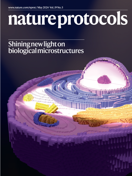
Illuminating digital cells on a microscopic stage.
A detailed digital reconstruction of an animal cell receiving light cast from afar. This represents the transformation of cells from microscopy images into a form suitable for accurate optical modeling of electromagnetic wave propagation.
Image: John Ball, National Eye Institute, National Institutes of Health. Adapted from Lauri Purhonen, Sketchfab, under a Creative Commons license CC BY 4.0. Cover design: S. Whitham.
This protocol describes a method for sampling the microbiome of food-processing facilities and analyzing it by using whole-metagenome sequencing. The protocol includes sampling and DNA-extraction and DNA-purification steps optimized for low-biomass samples.
FiLa biosensors allow real-time subcellular analysis of lactate metabolism. This protocol describes their preparation and characterization, and their use in in vitro and in vivo assays, under various nutritional and pharmacological conditions.
We provide a detailed roadmap for scientists interested in performing FDTD computational simulations to probe the interactions of electromagnetic waves (e.g., visible light or microwave radiation) with complex structures such as organs or biological cells.
The authors introduce MACHETE (molecular alteration of chromosomes with engineered tandem elements), a clustered regularly interspaced short palindromic repeats directed Cas9-based system for the efficient deletion of megabase-sized genomic regions.
Genetic interactions have been found to influence phenotypes in a variety of systems, yet their specific contribution to complex diseases remains unclear. This protocol describes Bridging Gene sets with Epistasis (BridGE), a computational approach for discovering interactions between biological pathways from genome-wide association studies data.
This multi-omics data integration protocol, which uses the web-based Analyst tool suite, covers knowledge-driven integration using biological networks and data-driven integration through joint dimensionality reduction.
Atomic force microscopy can be used to determine the stiffness of materials. This protocol describes how to measure and quantify the Young’s modulus E of pulmonary mouse and human basement membranes with atomic force microscopy and the Center for Applied Tissue Engineering and Regenerative Medicine processing toolbox.
Optimization of chemical transformations involves screening numerous reaction parameters. This protocol outlines how screening experiments can be rapidly and economically analyzed by quantitative benchtop 19F NMR spectroscopy.
We provide a twisting fabrication process for fiber electrodes that can be assembled into electronic threads and then integrated in electronic textile-based wearables.